Introduction:
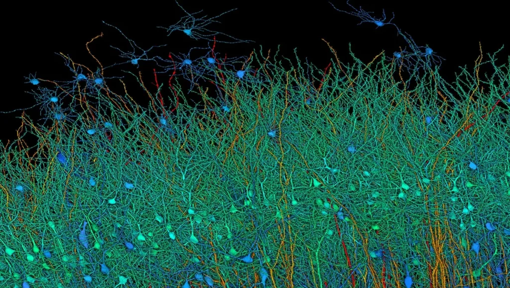
In a remarkable feat, researchers from Harvard University and Google AI have achieved an unprecedented milestone: creating a highly detailed 3D map of a tiny piece of the human brain. Google’s Brain imaging study captures intricate details down to individual neurons and synapses. It represents a monumental step forward in understanding the brain’s architecture and functionality.
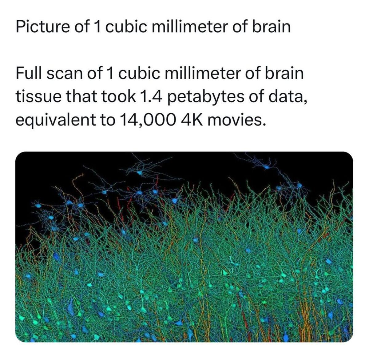
The study by Google shows that a mere 1 mm cube of human brain tissue yields approximately 1.4 petabytes of data. This highlights the monumental scale of data generation in neuroscience research. Integrating this data into storage infrastructure and computational capabilities offers profound insights into the challenges and advancements in the field.
Follow us on Linkedin for everything around Semiconductors & AI

Read More: 7 Reasons Why AI is the Future of Work – techovedas
Google’s Brain imaging study: Magnitude of Data
The sheer magnitude of the data generated from a single millimeter cube of brain tissue is staggering. With 1.4 petabytes of data, the scale of information captured is unprecedented. This amount surpasses the storage capacity of even the most advanced consumer hard drives by several orders of magnitude.
The researchers focused on a cubic millimeter of brain tissue—a minuscule volume that houses an astonishing number of components:
- 57,000 cells
- 150 million synapses
- 230 millimeters of blood vessels
Despite its small size, this cubic millimeter of brain tissue is incredibly complex, highlighting the immense challenge the researchers faced in capturing and reconstructing it.
Google’s Brain imaging study: The Mapping Process
To achieve such a high-resolution map, the team employed a meticulous process:
- Sectioning the Tissue: The brain tissue was sliced into 5,000 ultra-thin sections.
- Scanning with Electron Microscopy: These sections were then scanned using powerful electron microscopes to capture detailed images.
- Data Stitching with Machine Learning: Leveraging Google’s machine learning expertise, the images were stitched together to form a cohesive 3D image.
This process generated an enormous amount of data—1.4 petabytes, equivalent to roughly 14,000 4K movies. The sheer volume of data underscores the complexity and resolution of the map.
Read More: Top 5 VLSI Books for Beginners in VLSI Technology 2024 – techovedas
Google’s Brain imaging study: The Significance of the 3D Brain Map
This map is the most detailed representation of human brain tissue ever created. The map provides a revolutionary glimpse into the brain’s intricate wiring. It holds tremendous potential for advancing our understanding in several key areas:
- Neurological Diseases: Insights from the map could lead to better understanding and treatments for diseases like Alzheimer’s, Parkinson’s, and epilepsy.
- Intelligence and Learning: Understanding the structural basis of how our brains learn and remember could revolutionize educational methods and cognitive therapies.
- Brain Functionality: Detailed knowledge of neuron and synapse structures can shed light on how different brain regions communicate and function together.
Scaling Up:
Scaling this data to encompass the entirety of the human brain presents an even more awe-inspiring prospect. With the average human brain estimated to contain around 86 billion neurons, each forming thousands of connections with other neurons, the potential data volume becomes incomprehensible. Extrapolating to the entire brain could require storage capacities exceeding a zettabyte.
Read More: Beyond Nvidia: Top 5 Under-the-Radar AI Hardware Companies Poised to Take Off – techovedas
What’s the big deal?
The big deal about the 1.4 petabytes of data from this brain imaging study is two-fold:
Unprecedented view of brain structure: This is the most detailed map of the human brain ever made. It allows scientists to see individual neurons and their connections (synapses) in a way never before possible [2]. This is like having a blueprint of a tiny but crucial part of the brain’s circuitry.
Potential breakthroughs in neuroscience: By understanding how this millimeter of brain works, researchers hope to gain insights into how the entire brain functions. This could lead to major advancements in our understanding of things like:
- Neurological diseases: By comparing healthy brains to those with diseases like Alzheimer’s or Parkinson’s, scientists might be able to identify the root causes.
- Intelligence: This detailed look at brain structure could provide clues about how the brain processes information and gives rise to intelligence.
- Learning and memory: Understanding how the brain forms connections between neurons could help us understand how we learn and remember things.
Even though it’s just a small piece of the brain, it’s a giant leap forward in our ability to study this complex organ.
The Road Ahead
While this achievement is groundbreaking, it’s important to recognize that it represents only a tiny fraction of the human brain. The entire brain is about a million times larger than the scanned cubic millimeter. Scaling this project to map an entire human brain would require approximately 1.6 zettabytes of data—enough to fill a data center the size of 140 football fields. Currently, the technology and resources needed for such an endeavor are beyond our reach.
However, this study lays the foundation for future advancements. As technology continues to evolve, the prospect of mapping larger portions of the brain becomes more feasible. Each step brings us closer to unlocking the full mysteries of the human brain.


